The MRI technology has long established itself as an indispensable tool for medical diagnostics. However, today’s scientists continue to work on its development and the creation of new, improved systems. One of the most important aspects in this direction is the work with ultra-high-field tomographs, which can significantly increase the accuracy of medical research. Developers of the new MRI devices are trained at ITMO University’s Master’s program “Radiofrequency Systems and Devices”, which gives its students the option of choosing to study a specialized MRI track. While still in their Master’s studies, they can engage in scientific research, as well as participate in internships abroad as part of an international research group. More on the program’s educational process and what specialists it trains.
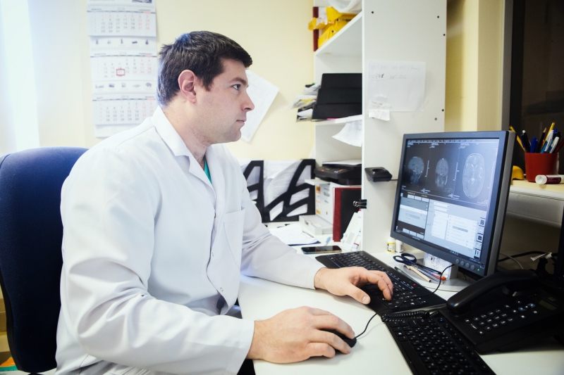
ITMO University’s Master’s program “Radiofrequency Systems and Devices” has been in implementation for two years. Its main aim is to provide Master’s students with in-depth knowledge in the field of radiophysics and, with that, the skills necessary for the development of real-world equipment and devices for modern radiofrequency communication networks, reception, transmission and processing of data, and systems for medical diagnostics.
Studying on the program, Master’s students can choose between two specialization tracks. The first, Radiofrequency Systems and Devices, is aimed at training the developers of antennae for modern communication systems. The second, MRI track, is connected to the development of radiofrequency systems for magnetic resonance imaging.
As emphasized by Stanislav Glybovsky, head of the educational program and member of ITMO’s Faculty of Physics and Engineering, the MRI track’s program was developed on the basis of a solid scientific foundation created at the International Research Center for Nanophotonics and Metamaterials.
We’ve created an international laboratory that conducts research in the field of MRI, and starting from 2014, we have been engaging in a range of research projects, including grants from the Russian Science Foundation and federal target programs on this topic. Owing to this work, we have been faced with a multitude of new tasks which created the need for preparing specialists with a similar focus, training students for carrying out research in the field of MRI methods and systems in general and the development of new radiofrequency coils, in particular,” comments Stanislav Glybovsky.
Demand for MRI specialists
All tomographs can be divided into several groups: low-field (with a constant field strength of up to 0.5T), medium-field (0.5-1T), high-field (up to 3T), and ultra-high-field (more than 3T). This division occurs due to their significantly different operating frequencies in the radio range, which are determined by the level of the magnetic field of a tomograph’s permanent magnet. Most modern clinics work with tomographs with a field level of 1.5 to 3T.
So far, ultra-high-field tomographs are only in use by high-scale research laboratories. For example, the largest European scientific centers such as Paris-based NeuroSpin carry out experiments on 7T tomographs with the capacity of containing the entire human body. In Russia, ultra-high-field MRI is conducted exclusively on special preclinical tomographs designed for use on laboratory animals. Working with these medical and research systems opens a wide range of new prospects and opportunities for the development of medical diagnostics.
First, an increase in the magnet field level brings a consecutive increase in the image resolution of the resulting image. Secondly, in addition to the quality of images, the field level also affects the speed of diagnostics. The higher the field strength, the quicker is the procedure. That way, an ultra-high-field tomograph with a field level of 7T has the potential to reduce the procedure’s timespan by several times compared with existing clinical tomographs.
As told by Stanislav Glybovsky, medical centers in France, the Netherlands and Germany already include entire departments of engineers and developers of radiofrequency equipment. They monitor the operation of tomographs, maintain the work of the devices, create radiofrequency coils and adjust tomographs’ settings for conducting different kinds of research.
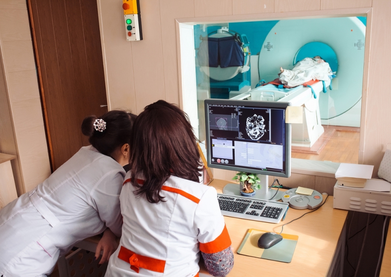
Why is this needed? Medical research centers carrying out research on ultra-high-field tomographs can’t operate on ready-made solutions, adds Stanislav. For example, various methods for studying parts of the human brain require special kinds of radiofrequency coils, many of which are not available on the market today. That’s why specialists who can independently develop a device for specific experiments are in very high demand.
“We regularly monitor job vacancies in the field and know dozens of European centers which show interest in specialists of this profile. But we’re also seeing some trends in Russian medical industry which give us reason to hope that in the near future, medical centers in our country which are currently only using ultra-high-floor tomographs for research on laboratory animals, will get access to 7T tomographs for conducting studies on the entire human body. That is why we want to train specialists who will be able to perform tasks like these,” comments Stanislav Glybovsky.
Competencies obtained by Master’s students
First and foremost, the MRI track provides students with a strong fundamental footing in the field of technical physics. But as noted by the head of the program, following successful training the program’s graduates not only acquire the competencies of a physics scientist but also the essential skills for an engineering developer.
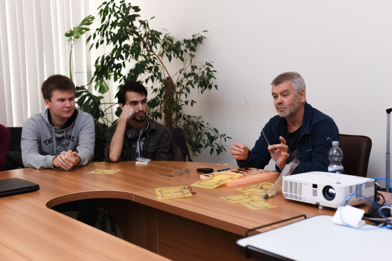
“Our goal is to train creative engineers. This means that graduating from our program, Master’s students should possess a profound understanding of the fundamentals of radiophysics so that in the future, they would be able to develop devices, for example, radiofrequency coils, designed for unique medical experiments. Why is this so necessary? The development of radiofrequency coils is a truly unique field; you can’t really expect to open a textbook and find a ready-made or even similar solution. Engineers working at a medical center face the tasks no-one solved before,” says Stanislav Glybovsky.
Education process within the MRI track
Choosing the track
Students choose their preferred tracks at the beginning of their studies. They are also given one month to decide on their scientific advisor. Тhis is important as it is this choice that defines a student’s scientific focuses and future field of work. But even having chosen one track, you can add on the subjects studied as part of another one if you so wish.
Basic training
Because students from different universities join the Master’s program, at the beginning of their studies they are offered subjects aimed at helping them revise on the information needed to proceed further into the course. Among such subjects are technical electrodynamics, radio circuits and signals, and quantum radiophysics. These are the basic courses that lead up to more advanced studies of the special sections on the development of radiofrequency coils, as well as magnetic nuclear resonance methods for tomography and spectroscopy.
Research work
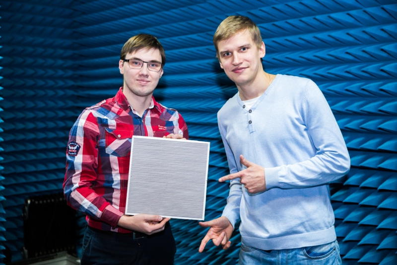
Research work begins as early as in the first year of the Master’s program, and this is the key drawcard of its education process. An entire range of projects in the field of MRI are currently being pursued at ITMO’s Faculty of Physics and Engineering, and students who from the beginning of their studies have expressed interest in scientific research can join this work. Candidates who have successfully passed the probation period are enrolled as regular staff and allocated a salary.
Courses from leading specialists in the field
As noted by Mikhail Zubkov, a research member of the Faculty of Physics and Engineering coordinating the education process within the MRI track, the faculty’s staff are actively cooperating with several international research groups recognized as leaders in the field of the development of radiofrequency coils for MRI. They include the research groups of Prof. Andrew Webb (Leids Universitair Medisch Centrum, the Netherlands), Dr. Nico van den Berg (University Medical Center Utrecht, the Netherlands), as well as scientists from CEA Neurospin (Paris, France), Max Planck Institute (Tübingen, Germany), Center for Genetic Engineering and Biotechnology (Havana, Cuba), as well as Center for Advanced Imaging Innovation and Research in New York.
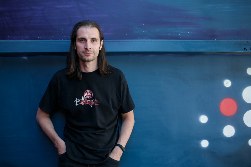
The scientists’ collaboration consists not only in solving joint research tasks but also in exchanging experience. For example, ITMO has recently welcomed Nikolai Avdievitch, a senior research associate at the Max Planck Institute in Tübingen (Germany), who taught a special course for the university’s students. In the framework of the program, the scientist spoke about his work and presented the modern techniques used for the development and manufacturing of new radiofrequency coils and electronic devices for experimental ultra-high-field MRI (you can read the full ITMO.NEWS interview with Nikolai Avdievitch here).
“We are glad that our scientific contacts allow us to not only conduct joint research but also help us invite leading specialists in the field so that they could share their knowledge with our students: to give a series of lectures or hold workshops,” comments Mikhail Zubkov. “Apart from that, from their very first year into the program, our students can participate in research internships at our partner institutions to absorb their expertise in the course of joint work.”
Internships and work as part of international research groups
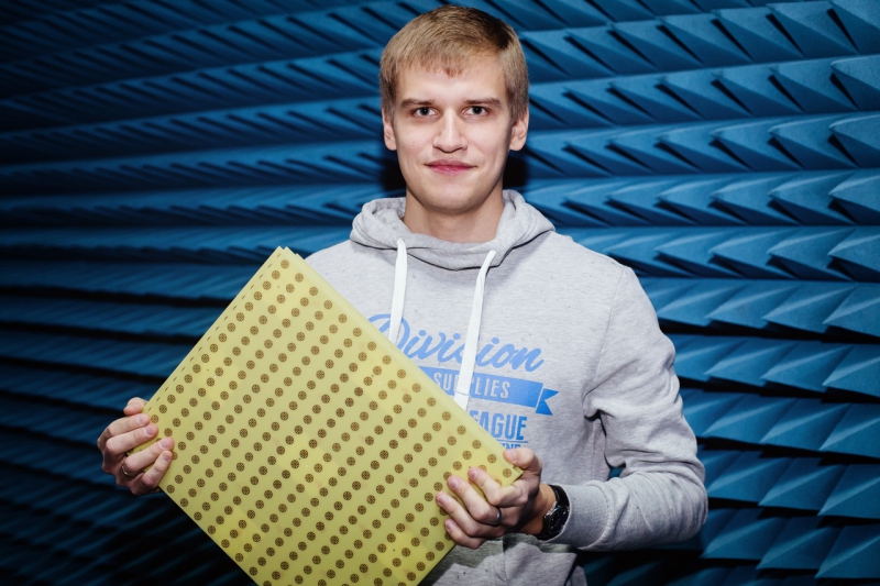
As one example, last year Ksenia Lezhennikova, a second-year student of the program, did an internship at the Center for Magnetic Resonance in Biology and Medicine of Aix Marseille University (AMU), where she could familiarize herself with the work of a 7T ultra-high-field tomograph and conduct research into developing her own radiofrequency coil.
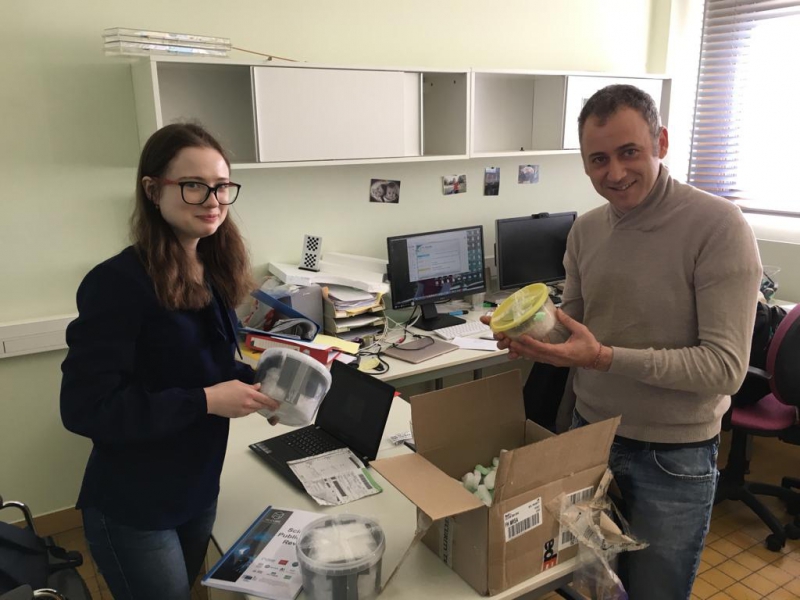
“I’m working on the project Magnetic Screen, which is part of a larger program M-Cube. In the course of our work, after we have completed the modeling stage, it became necessary for us to conduct relevant experiments and assemble a sample of our coil and test it on a tomograph. To do this, we needed to come to France to visit our colleagues with whom we cooperate. While at Aix-Marseille University, I did a one-month work on developing my sample. I also had the opportunity to attend an experiment that made use of a real tomograph to see how other coils are tested. It was a very rewarding experience. As early as in the first year of Master’s program, I had the chance to go to a university in another country, work with my international colleagues in the field, and attend a scanning procedure that involved an ultra-high-field tomograph, and this is something not many people get to do,” says Ksenia.
What is more, after her trip to France, Ksenia also managed to participate in a summer internship at Aalto University in Finland. She is now continuing her research in the field of MRI and plans to pursue scientific activities later in her PhD studies.
The program’s graduates can continue their research career both on the basis of ITMO University’s PhD studies or abroad in Europe, adds Stanislav Glybovsky. For example, one of the Master’s program alums is now conducting his research at Institut Langevin and NeuroSpin in Paris, while other graduates continue collaborating with ITMO’s European partners as PhD students.
Elena Menshikova
Journalist
Anastasia Krasilnikova
Translator
